Artificially made hair implants
Preface
Dear readers,
As you might known from the media, people are invoked with various information campaigns to get involved as an organ donor, since the number of available and the acceptance of donor organs through the recipient’s immune system is often not sufficient to provide all people who need an organ transplant with transplants. For this reason, efforts have been made for some time to develop new sources of transplantable organs. For example, it is not unusual today for patients who require a new heart valve to receive those derived from a pig heart. However, transplantation medicine has narrow limits across species.
For this reason, attempts have been made in the laboratory to produce artificial tissue and organs, what is called bio- or tissue engineering . These include the advantage that there will be no rejection reactions and the graft will be fully accepted by the recipient’s immune system. In addition to the possible production of kidneys, heart valves or livers, these techniques would also be interesting for people who suffer from alopecia, but can not use the possibilities of a hair transplantation due to a too small donor area. Here exists the idea that the hair follicles needed for such a treatment can be produced artificially in a cell culture.
In fact, such considerations arose since the 1970s and since this time are attempts made to realize these ideas. But what is the biomedical state of artificial hair transplant creation currently? In my new article, I would like to explain the current state of research and its basics. In the following, not only the question of how patient-specific hair transplants could be generated will be addressed, but also will be discussed what the actual obstacles are in the investigation of such treatment options, and what realistically can be expected in the coming years from biomedical research.
Therefore, enjoy reading!
Sincerely
Yours, Angela Lehmann
The Artificial Production of Organs and Tissues as Potential Source of Hair Transplants
In the past years so-called bio-engineering or tissue engineering – the artificial production of organs and tissues – has made spectacular progress. There are always reports that in the near future even entire organs could be produced in the laboratory since implants are urgently needed – with maximum immunocompatibility and tailored specifically to each recipient. Comparable efforts are made to create recipient-specific hair transplants. For this reason I would like to inform you in my new article about the current state of research investigating hair transplants from the cell culture laboratory.
How does a cell know what its job is in the body?
Before we can approach the question of how to create artificial tissues and organs that could potentially lead to a revolution in transplantation medicine, we need to take a look at the question why a somatic cell “knows” what its specific task is in a particular organ or tissue. Here you can first imagine a fertilized human egg. It divides itself first, what results in two identical cells. This process of division happens again and again, so that the number of cells increases steadily. At some point, however, the cells undergo a change, which means, they undergo the process of specialization. While in principle a fertilized human egg can become every other cell in the body, such as a skin, liver or nerve cell, already specialized cells can not “choose” another specialization. This process is also referred to as cellular differentiation and is naturally irreversible in mature somatic cells. This means that a differentiated nerve cell will always remain a nerve cell.
The differentiated cells are the reason why the body is composed of different tissues, the term “tissue” always refers to a aggregation of specialized cells of the same type. Different tissues differ since their cells have characteristic gene expression profiles. This means, for example, that in a liver cell more genes are expressed, which are related to the detoxification of the body, while in nerve cells more genes are expressed, which have something to do with the nervous information forwarding. Furthermore, the cell types differ from each other so that the cells of one type divide faster than another. Among other things, this results in the fact that different tissues can regenerate at different rates and quickly. Accordingly, injuries to the skin usually heal within a few days, while it may take years for nerve tissue to regenerate (if at all) after a stroke, for example.
This remaining ability of cellular differentiation is referred to by various terms. For example, while a fertilized human egg can make up every other cell in the body (this human egg cell is called totipotent), certain stem cells can only produce particular tissues (referred to as pluripotent or multipotent cells). Nonetheless, in recent years it has been discovered that pluripotent stem cells are also present in the organs and tissues of an adult human, which potentially can be used for the therapy of certain diseases because of the fact that they can still form many other tissues. On the other hand, if a once specialized cell no longer divides, but “merely performs its task” in the body, it is called a senescent cells.
It should be mentioned at this point that one of the challenges to be discussed below is that an organ does not always consist of only one tissue, but of cell aggregates of different types. For example, human skin differentiates between several types: fibroblasts are the precursors of connective tissue cells, whereas keratinocytes, for example, are involved in the formation of the epidermis, whereas melanocytes are responsible for the color of the skin through the production of pigments. Accordingly, even a hair follicle can not be called a tissue, because in fact it is a hair organ.
How cell cultures could be influenced to produce specific tissues and organs?
All methods designed to produce organs or tissues in the laboratory now aim at modifying this cellular evolutionary specialization program. Numerous approaches are available for this purpose. But here too, a brief historical digression must be made first:
By the middle to the end of the last century, most researchers were still convinced that an adult body contains only very few pluripotent, tissue-forming stem cells that could be used for therapy to only a limited extent. Such cells have been known, for example, in the bone marrow or hematopoietic system. At that time, among other things, it was also believed that the nerve cells of the brain had no longer any regenerative potentials, so these cells are senescent and present, but only fulfill their biological tasks. However, over the past 30 years, more and more tissues of the human body have been identified that show a stem cell activity even in adulthood that could be used as a source of tissue regeneration. While these are not totipotent stem cells (those that could all form tissues), adult stem cells are still capable of forming many related tissues. Thus, adult neuronal stem cells can still differentiate into nerve cells (neurons) and microglia (these ensure the nutrition supply of the neurons), which is important for the regeneration of the brain, for example, in cases of stroke.
In order to be able to obtain stem cells of whatever kind for the bio-engineering or tissue engineering of transplants, a first step is to isolate them from the body. This is necessary due to two reasons: First, the stem cells often do not occur in the body exactly where they are needed for therapeutic intervention (for example, certain stem cells are at the bottom of the hair follicle, whereas they would be needed for therapeutic use in the overlying cell layers). On the other hand, the number of adult stem cells in the body is usually too low to use these cells for therapy. Isolation now gives rise to the possibility of bringing these stem cells outside the body into nutrient solutions with corresponding growth and differentiation factors, causing them to multiply (the ability to divide can sometimes be greatly stimulated outside the body) and to differentiate into a desired cell type. Sometimes multiplication is termed “cloning”, but this term is problematic in the given context. Here, however, the first problems arise: Stem cells outside the body in a culture dish (Petri dish) behave not as they do within the body. For example, the cultures of the stem cells lose their ability to divide or regrow without any apparent reason, spontaneously differentiating into an undesired cell type appears or, over time, cells become hybrid cultures that may not be of interest for further investigation. It is therefore a great challenge to find the right culture conditions for each individual cell type. This often happens even today according to the principle of trial and error.
Once it is possible to determine the correct culture conditions, it is still important to give the cells the right signals for a possible differentiation. There are numerous growth factors available for this, which can cause a variety of effects. Again, the right combination must be tested in order to achieve a satisfactory result. However, the cultured stem cells are also influenced by effects that are difficult or impossible to reproduce in the laboratory. For example, for successful differentiation, contacts between cells of the same type but also between different types are needed. In this way, the cultured cells communicate with each other, exchange signal substances or arrange themselves in a certain way in the culture dish. If one cell type is stimulated by another to differentiate, the term induction is used.
In order to make clear that the testing of growing conditions is not trivial, the development of inducting new hair follicles is presented: Induction of hair follicle formation was first demonstrated in rodents in 1970. These animals show an inductive potential of papillary cells (cells of a skin layer). However, it took 14 years to test the right culture conditions, so that this inductive potential was retained even in cell culture. However, the researchers had great difficulties transferring this knowledge to the human organism. In 1999 it was possible to prove the inductive potential for hair follicle formation with human sheath cells (which are located in the skin just below the epidermis) implanted in an intact skin surface. Noteworthy, however, is the fact that this transplantation was carried out across the gender barrier in humans and was nevertheless successful. Again, it took until 2012 before it was possible for the first time to produce an entire hair organ in a recipient body by stem cell transplantation. This new hair organ was connected to the complete supply of nutrients (blood and lymph circulation) of the surrounding tissue, the new hair follicles went through several hair cycles and the formed hair could raise through an existing hair muscle (piloerection, in humans termed goose bumps). However, rodents were again used as experimental system for this experiment and until today it was not completely possible to transfer these results to the human organism. The reason for this is still a mystery to the researchers – but they work on that problem.
What is the current situation regarding the production of hair follicles for hair transplantation?
Regarding the human organism, the unequivocal identification of the pluripotent stem cells in the skin, which can form nearly all the tissues of the skin, was a spectacular finding of recent research. By a lineage analysis, these cells, which can differentiate into all skin cells (including the hair organ), could be identified in the body. These stem cells are located at the bottom of a hair follicle invagination, and hair follicle formation can be induced by contact with partially specialized skin cells. A little later, it was possible to show that this ability is maintained even when the cells are isolated and propagated in cell culture.
However, at this point, the researchers encountered another problem that was not initially in the focus of interest: Organs, and thus the hair organ, are three-dimensional objects that can only be produced insufficiently in a flat, two-dimensional cell culture dish. This is due to the fact that the optimal growth or differentiation conditions are not yet guarantors that the cells spatially arrange themselves as it would be the case with a three-dimensional hair follicle in the skin of a human being. For this purpose, further modifications of the experimental conditions are required.
Studies investigating the three-dimensional growth of cell cultures led to a breakthrough in 2013: It was known at that time that human papillary cells have a inductive potential to stimulate stem cells to form new hair follicles. But this ability decreases in cell culture which has been attributed to the lack of cellular contact with surrounding tissues, such in the epidermis (upper layer of the skin). Based on these considerations, the researchers proceeded to use a three-dimensional structure instead of a two-dimensional experimental design. In this three-dimensional design the cells did not grow on the bottom of a cell culture dish as before, but floated freely in droplets on the ceiling of a Petri dish. The following figure illustrates this difference in growth conditions.
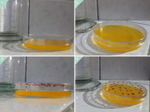
The figure shows the difference between a two-dimensional and a three-dimensional cell culture. While it is visible that in the two-dimensional variant both the (1) side view and the (2) top view show that the cells form only one layer at the bottom of the Petri dish, the cells organize themselves in the three-dimensional experimental arrangement in the (3) side view and the (4) top view on the ceiling of the Petri dish in hanging drops of the nutrient solution.
Even if one could believe that this represents only a very small change in the experimental conditions, this approach gave rise to very interesting changes: In studies concerning the gene expression profiles, i.e. the question of how strongly certain genes are expressed in the cell, it could be asserted that the three-dimensional cell cultures are 22% more similar to the desired hair follicle stem cells than the cells grown in a two-dimensional cell culture. These cells from a three-dimensional culture were subsequently transplanted to hairless mice and new hair follicles formed that consisted of both human and mouse cells and produced hair. Further investigations must subsequently show whether this mechanism also works in a human-to-human transplantation. However, this experiment remains groundbreaking as it has for the first time been possible to isolate and cultivate human skin stem cells without losing the ability to form hair follicles and this has then been demonstrated by transplantation to another organism.
Nevertheless, research about the bio-engineering of hair follicles (also referred to as hair follicle cloning), which would be useful for the treatment of alopecia or other forms of hair loss, still poses many difficulties. Of these, the identification of correct culture conditions and the change from a two- to a three-dimensional experimental design have already been discussed. Other challenges are that the newly formed hairs do not always have the desired texture. Thus, the hairs formed are sometimes significantly thinner or thicker or have no color, since the pigment-forming melanocytes are missing in this experimental approach. A further problem still results from the spatial arrangement of the cells: Although hair follicles form after a transplantation, they grow together, are generally not sharply demarcated, or several hair shafts grow from one follicle. These are then disordered, so that the later growth direction of the hair in the case of a hair transplantation would be difficult to determine if one would use them as micro grafts. It is still unclear how many hair cycles such a follicle can go through before it stops producing new hair. Without this information, it would be difficult to estimate how sustainable a hair transplantation would be by means of artificially produced hair follicles. In addition, it must not be forgotten, that such research is always associated with a great financial costs and that financial interests always influence the researchers and their institutions.
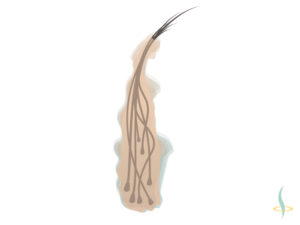
The figure shows how several hair shafts grow in different directions from one artificially produced hair follicle.
In conclusion, research on bio-engineering a hair follicle or hair follicle cloning is certainly well on its way, as the successes of recent years have shown. However, there is much more work to be done, so that the treatment of hair loss by artificially produced hair follicles will not be possible in the next days, but it can be expected that in the coming years and decades the procedures can be perfected to provide a widespread therapeutic use of laboratory-produced hair follicles.
However, hair transplantation is already a tried-and-true procedure that allows people suffering from alopecia or other forms of hair loss to counteract their condition. Whether this is a suitable therapy option especially for you and whether, for example, the donor area is sufficient for a treatment, can not be said in general. This requires expert advice and education of patients who are interested in hair transplantation. If you also feel uncomfortable due to increased hair loss, or are interested in the possibilities and limitations of hair transplantation, I would like to encourage you to contact us for a consultation in our clinic to find a responsible solution.
Sincerely
Yours, Angela Lehmann
References
Higgins, C. A., Chen, J. C., Cerise, J. E., Jahoda, C. A. & Christiano, A. M. (2013). Microenvironmental reprogramming by three-dimensional culture enables dermal papilla cells to induce de novo human hair-follicle growth. Proceedings of the National Academy of Sciences, 110(49), 19679–19688.
Stenn, K. S. & Cotsarelis, G. (2005). Bioengineering the hair follicle: fringe benefits of stem cell technology. Current opinion in biotechnology, 16(5), 493–497.
Tezuka, K., Toyoshima, K. E. & Tsuji, T. (2016). Hair follicle regeneration by transplantation of a bioengineered hair follicle germ. Multipotent Stem Cells of the Hair Follicle: Methods and Protocols, 71–84.